


Functional MRI (fMRI)
The Challenge
The use of functional MRI (fMRI) in neurological research and practice has grown dramatically. This important tool is uncovering many of the mysteries of the brain and its response to diseases, stimulation and injury. Learning and imaging the underlying brain response to pain is impossible though, without pain stimulator that can work in an imaging environment.
The Science of Imaging Pain
The anterior cingulate cortex (ACC) has been associated with many aspects of behavior, including pain. PET studies have consistently shown ACC activations during noxious thermal tasks. fMRI studies of individual subjects indicate that a small region of the posterior aspect of ACC can be activated during painful median nerve stimulation and is separate from the more anterior ACC region involved in attention.
Studies using fMRI with the PATHWAY stimulators can help determine the spatial distribution of cool, warm, cold pain, heat pain and motor-
Applying Medoc Solutions in fMRI
Medoc’s PATHWAY, non-
In this application the PATHWAY model CHEPS and ATS are not used as a diagnostic tools, but rather sophisticated pain stimulators integrated in the fMRI system and synchronized with its main computer. Medoc’s stimulators are currently being used by pain and neurological researchers at the Harvard University Medical School, the University of Toronto, pain research institutes in England, the University of Munich, the University of Birmingham in Alabama, the Veterans General Hospital, and National Yang-
Medoc fMRI Components & Configuration
- PATHWAY model CHEPS & ATS
- Special non-
magnetic 30x30mm or 16x16mm Thermodes for TSA or 27mm diameter Thermode for CHEPS, with 10 meter long cable - TTL Output for triggering external device
- TTL Input for triggering the PATHWAY from external device
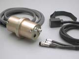
- 70 mm fMRI diameter filter for the thermode
- Non magnetic CoVAS
- Non magnetic patient response unit and emergency button
The stimulator is located outside the MRI room. The special thermode adapted for use in a magnetic environment is inserted into the room via a special hole in the wall and through the filter.
* THE PATHWAY WAS SUCCESSFULLY TESTED TO WORK WITH UP TO 4 TESLA MRI SYSTEMS.

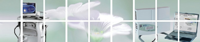
Ramp & Hold screen
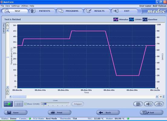
Run Test Screen
click on the images to enlarge
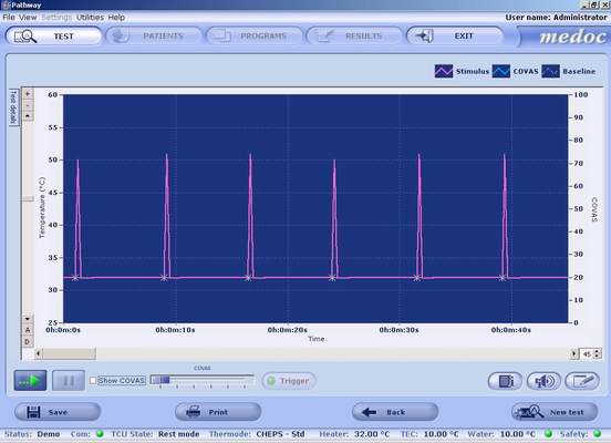
Pathway System
fMRI filter
Pain Research applications: